Faculty Spotlight: C. Forbes Dewey Jr.
A Lifetime of Bioengineering Achievement
by Alissa Mallinson

Photo credit: Tony Pulsone
Professor of Mechanical Engineering and Biological Engineering C. Forbes Dewey Jr. first came to MIT’s Department of Mechanical Engineering in 1968 as an associate professor, bringing with him a BS in mechanical engineering from Yale University, an MS in mechanical engineering from Stanford University, and a PhD in aeronautics from California Institute of Technology. Between his PhD at Caltech and his position at MIT, he did a five-year stint at the University of Colorado and the Joint Institute for Laboratory Astrophysics in Boulder, where he struggled to tame unstable plasmas in the hopes of achieving controlled fusion.
“Back in 1968,” remembers Dewey, “there were a few very forward-looking faculty members here at MIT, including Professor Ascher Shapiro, who believed that bioengineering was a great new field that was going to integrate biology, chemistry, physics, and engineering, and transition into something miraculous. I thought that was very interesting.”
With a background in fluid mechanics, Dewey was a perfect fit for this great new field. The number of engineers studying biomedical fluid mechanics at the time was miniscule, and many very challenging problems were being discussed. “I said to myself, ‘I’m an engineer, this is biology. How can I make a difference?’
“But then I got to thinking about blood flow, as exemplified by the flow past an atherosclerotic artery constriction, and the question of where the sound comes from was a very interesting physics problem to me,” he recalls. “You have blood flow through a restricted artery, and you should be able to at least get a theory for what that looks like and try to understand where the sounds come from.”
He eventually transitioned from fluid-mechanical problems to the cells themselves in order to study atherosclerosis. At branch points, arteries are more prone to developing disease. Dewey knew it was a function of their geometry and blood flow, and suspected that the underlying mechanics of the problem were related. But he quickly realized that external engineering studies couldn’t get to the answers he was seeking, because the key phenomena were all biological in nature, and that he’d have to find a way to get a closer look.
Partners in Discovery
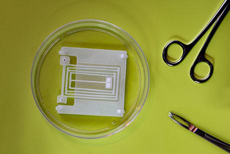
Dewey partnered with Dr. Michael Gimbrone, a pathologist at Brigham and Women’s Hospital who had developed new techniques for growing cells in culture. Together they designed a laboratory apparatus that would first grow the cells, then subject them to fluid flow, mimicking what they would experience in the artery. The apparatus was modeled after a cone-and-plate viscometer, well known to the fluid mechanics field for producing a constant shear force over a large surface area. This allowed large samples of cells to be exposed to shear and make them available to biological assays.
The cone-plate apparatus allowed Dewey and Gimbrone to make the first controlled in vitro laboratory measurements of the effects of fluid flow on vascular endothelial cells. Today there are a number of different apparatuses to perform such experiments, and the response of cells to fluid shear stress is studied in hundreds of laboratories around the world, but at the time, it was a groundbreaking invention.
Now with the ability to visualize blood flow, Dewey and Gimbrone began to look closely at the cell mechanics. They were able to look at the interior of the cells to see how the force of the blood flow not only changes the cells’ orientation to align with the flow direction, but also changes the arrangement of the internal structural members in the cell and brings about dramatic changes in cell crawling speed and morphology. The endothelial cells that were subjected to disturbed flow [flow that oscillated and had a low magnitude] became compromised, shedding from the surface and leaking at the joints between the cells. Dewey and Gimbrone realized that they were likely visualizing the very same phenomena that occurred in vivo in the very regions of bifurcations subjected to disturbed flow.
In 1988, Dewey ran into a problem. “Using various methods, we went looking for the actual mechanism that couples the fluid force to the cell, and came up with a blank,” he says. He felt that early theories explaining that shear stress acted directly on the cell membrane were inadequate. How could a “fluid membrane” resist a continual force without being totally distorted, he thought? Frustrated, he turned his attention to other interesting problems, becoming heavily involved in the development of medical imaging standards, such as the DICOM standard, as well as nonlinear optics. He and Lon Hocker developed novel devices for broadly tunable wavelength laser systems and, with the help of MechE Professor Roger Kamm, pollution device detection systems based on resonant opto-acoustic spectroscopy.
There and Back Again
With such life-changing discoveries already under his belt, Dewey could have easily stopped there. But instead he returned to the mystery of the fluid-force-cell connector. He ran across a paper published by Professor Brian Duling of the University of Virginia showing that on top of the cell membrane is a very thin layer of gooey material about 0.5 microns thick called the glycocalyx.
“All of a sudden, that layer became all important to me,” says Dewey. “Now you had your mechanism for getting the force from the fluid flow to the inside of the cell.” With this missing piece of the puzzle finally in place, Dewey’s research team was the first to directly measure the mechanical stiffness of glycocalyx in an ongoing and as-yet unpublished experiment using super-resolution microscopy and a quantum dot florescence technique.
“It all made perfect sense,” says Dewey, “and for the first time it was possible to visualize how the shear stress actually acted on the glycocalyx and its support structure, which reached completely through the cell membrane and attached to the cytoskeleton. It was also clear that if the glycocalyx was missing, the effects of fluid shear stress on the cells would be compromised. The potential for understanding how fluid flow and cell biology worked together to modulate arterial disease seems at hand.”
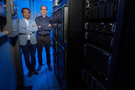
Just as he did in earlier years, Dewey is simultaneously working on data collection and analysis as well – specifically, highly complex models for characterizing drugs for development. Back in 1981, Dewey was a founder of the Massachusetts Computer Corporation, a successful venture to develop microprocessor-based laboratory computers for science and engineering. That love for computational problems was triggered in Dewey again when it became obvious that the drug development process was eating larger and larger chunks of the health care budget, with poor results.
“Many people have created elegant small- and mid-scale models for drug characterization, but they eventually become so large and complex that they are impossible to deal with,” he says. “There are too many species and reactions, and no simple way to modify the data when new information comes to light. So what we’ve done is to treat each of these individual pathways for combination as a black box, characterize the black box, and then program the computers to put the black boxes together.” A desire to solve the problem led Dewey to found another new company, CytoSolve, Inc., with a former student, Shiva Ayyadurai.
“The challenge of making a positive impact on increasing the success of drug development – helping many people with cancer and cardiovascular disease lead better lives because of more intelligent drug design – is very exciting.”
Professor Dewey is a founding fellow of the American Institute of Medical and Biological Engineering, an inaugural fellow of the Biomedical Engineering Society, a fellow of the American Physical Society, and a foreign fellow of the Royal College of Engineering (UK). He is also a vice president and trustee of the Fidelity Nonprofit Management Foundation.