New approach uses computer vision to assess children with cerebral palsy for optimal treatment paths
Clinical gait assessment collected using cell phone video reduces existing barriers to treatment
Cerebral palsy (CP), a neurological disorder caused by atypical brain development or brain damage, is a common childhood motor disability that affects movement, muscle tone, and posture. The condition usually presents early in life and can have symptoms ranging from mild to severe. While there is no cure, therapies can improve function for many patients.
“A child with Cerebral Palsy needs regular assessment,” explains Eran Beeri Bamani, a postdoctoral associate in the Department of Mechanical Engineering (MechE) at MIT. “Usually, this requires the patient to go to a hospital and be observed by clinicians.”
During assessments patients are given a clinical score with a label of one through five. “Level one is the beginning of the disease, level three can barely walk, level four cannot walk,” Bamani explains. The assessment, and the accuracy of the label, are essential for treatment, but current practices to collect this information are limited by factors like data scarcity, access to clinicians, class imbalance, and by the clinical diversity present among patients.
Bamani and fellow MIT MechE Postdoctoral Associate Joao Buzzatto are first authors on a new paper that that presents an approach using computer vision to classify children with cerebral palsy. The integrated artificial intelligence framework, which is designed to automate the assessment of gross motor function through standard smartphone videos, seeks to remove some of the barriers clinicians face in making assessments. Hermano Igo Krebs, principal research scientist in MechE, is also one of the paper’s authors.
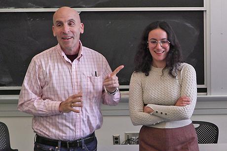
 Postdoctoral Associate Eran Beeri Bamani demonstrates an approach using computer vision to classify children with cerebral palsy. Credit: Tony Pulsone, MIT Mechanical Engineering
Research in Krebs’ lab, The 77 Lab, sits at the crossroads of mechanical engineering and design, computer science and control, and neuroscience and human factors, pushing human-robot interactions to empower humans. The idea behind this paper came from something the team has been working on for quite some time. “We are looking to ways to create biomarkers, and to use robots to deliver therapy, but at the same time, evaluate patients based on how they make their movements,” says Krebs.
The group’s past projects required the patient to be present, on site, with the robot for evaluations to occur – the team saw a better way. While studying another disease, metachromatic leukodystrophy, which is a rare and fatal condition that also requires patients to make regular visits to care teams for assessment, the team began to ask what they could do to make the process easier for the patient and for caregivers.
“Everybody has a smart phone,” says Krebs. “The idea came that we could use the smartphone to take videos of the patients and, from these videos, we could extract the clinical scores.”
The team started with a gross motor function scale for patients with metachromatic leukodystrophy, then progressed to CP for a broader dataset that would allow them to train their AI models more effectively.
They employed a Conditional Skeleton Diffusion Model (CSDM) to generate anatomically accurate synthetic gait sequences using time-series coordinates of human joints to analyze motion. Converting the data to skeletons helps to preserve privacy. Sequence samples are then combined with real patient data to train the model and create an index score. The approach offers a scalable solution for continuous clinical monitoring in home settings and was found to reduce errors and improve recognition of severe phenotypes. In this study, the model achieves a high classification accuracy of 85.7 percent.
The study demonstrates that generative data augmentation significantly improves the recognition of rare and severe motor phenotypes, providing a more robust tool for patient treatment and rehabilitation. Further, the researchers say this approach could be used for assessment in conditions beyond metachromatic leukodystrophy and CP, including Parkinson’s or for the treatment of patients with brain tumors.
Krebs says it’s exciting to see the results and the direction this work is heading. “You can calculate everything in the smartphone,” he says. “Data privacy, all these aspects, are less of a concern… we can give results and eventually even use the phone to deliver therapy.”
The paper, “Diffusion-Augmented Spatio-Temporal Graph Convolution for Clinical Gait and Motor Function Assessment” is available now from IEEE-Transaction of Neural System and Rehabilitation Engineering. Funding for this study was provided by U.S Department of War: U.S. Army Medical Research Acquisition Activity (USAMRAA).