MIT Researchers develop new way to explore T cell exhaustion
Research offers new way to study immune response
T cells (or T lymphocytes) are a type of white blood cell that plays a central role in the immune system, destroying infected or abnormal cells and coordinating the overall immune response. Chronic infections, such as Tuberculosis, HIV, malaria, or cancer can persistently stimulate a body’s T cells, pushing them toward exhaustion, which can impair their protective function.
This exhaustion has widespread implications for the progression and treatment of chronic diseases, but current detection methods require expensive and tedious antibody labeling, destructive workflows, or days-long functional assays that limit dynamic monitoring capabilities. Researchers in Assistant Professor Loza Tadesse’s lab wondered if it would be possible to identify T cells’ exhausted traits at high throughput without labeling or damaging the cells, which would allow new ways to explore supporting the immune system.
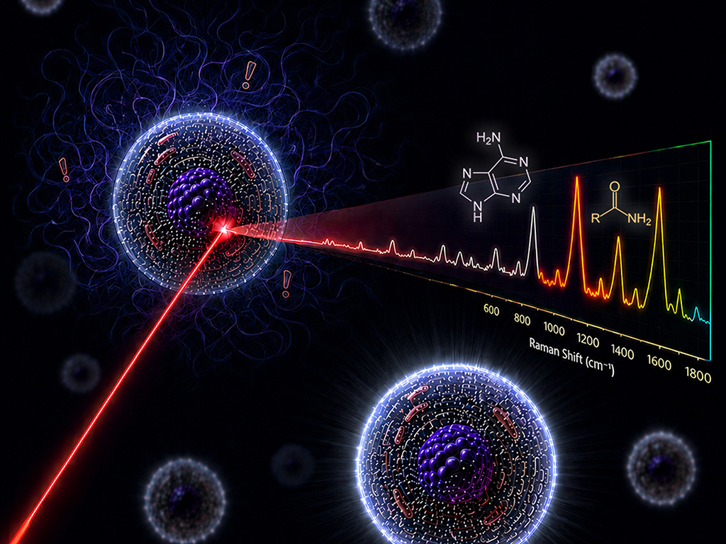 T cells can be identified within seconds through their intrinsic Raman spectral fingerprints. Molecular vibrations associated with adenine and amide, depicted above the spectrum, illustrate key biochemical features that contribute to high spectral classification accuracy. Image credit: Image generated by ChatGPT-5.6 Sol, with subsequent edits by the authors. Courtesy of the Researchers.
“This may be far off, but perhaps one day we could give exhausted cells a “rest season” and return them to the fight if their function recovers,” says Tadesse, who is the Latham Family Career Development Professorship. “In the here and now, a non-invasive way of reading immune-cell states could help us continuously monitor how the presence of pathogens, added antibiotics and other tissue can affect the immune response and infection resolution.”
In a paper recently published in the journal ACS Sensors, Tadesse’s team demonstrates an intelligent Raman spectroscopy-based label-free non-invasive approach to telling T-cell activation and exhaustion. MIT authors include Tadesse, Marissa Morales (lead author, HST), Srilakshmi Premachandran (MechE), and Shruthi Ravichandran (EECS).
“T cell exhaustion has been shown to influence treatment response to some cancer therapies and even predict the development of HIV-associated cancers months before a diagnosis,” says Morales. “Our goal was always to try and enable faster access to this information about T cell state so that it could one day more readily inform clinical decision making."
The team studied healthy donor-derived T cells that were suspended in media and systematically stimulated to model exhaustion, then distinguished unstimulated, activated, and exhausted cells with over 97% accuracy across three donors and multiple spectroscopy hardware setups.
The “gold standard” for cell sorting requires costly, time-consuming antibody staining to measure surface markers. In contrast, Raman-based workflow acquires spectra in seconds. Tadesse says this work opens a new window for her lab research into infection.
“We have long focused on identifying disease-causing pathogens; now, we can begin exploring the other side of the battle, the immune response,” Tadesse says. “This could help us follow interactions between immune cells and bacteria in real time and investigate how antibiotics change those dynamics. Furthermore, when identifying the pathogen is a needle in a haystack problem, the immune response may offer us clues to disease stage and prognosis.”
The paper, “Nucleic Acid Turnover and Lipid Remodeling Distinguish T Cell Activation and Exhaustion States via Label-Free Raman Spectroscopy,” is available now from the journal ACS Sensors.